"You have posited an very important hypothesis.
Clearly, you have made a good argument
for a central role of cholesterol homeoststis in
AD pathogenesis."
"Thank you for your most interesting communication."
(expert readers feedback)
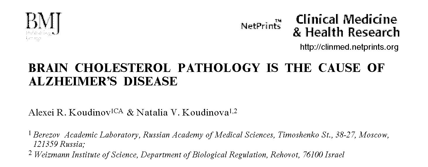
Brain Cholesterol Pathology is the Cause of Alzheimer's
Disease.
Koudinov AR, Koudinova NV
Clin Med Health Res (2001) Published online November
27, 2001. clinmed/2001100005
[ Current article Hit Parade: 2356
readers ]
Abstract
Several recent reports provided important knowledge on how inhibiting
cholesterol production in the brain might inhibit amyloid b(Ab)
production, and reduce the accumulation of Ab
that causes Alzheimers disease (AD). As we show and discuss here cholesterol
homeostasis biological misregulation itself has a key role for synaptic
plasticity impairment, neuronal degeneration and is the primary cause for
several AD hallmarks not limited to brain amyloid. Moreover, Alzheimer's
changes in neurochemistry of Ab, tau, neuronal
cytoskeleton, and oxidative stress reactions likely represent physiological
transitory mechanisms aiming to compensate impaired brain cholesterol dynamics
and/or associated neurotransmission and synaptic plasticity failure.
Authors key words:
Alzheimer's disease, amyloid beta precursor, cytoskeleton, Down syndrome,
learning, memory, lipoprotein receptor, LTP, neurodegeneration marker,
oxidative stress, PHF tau phosphorylation, plaque, phospholipids, synaptic
plasticity, secretase, SREBP
   

"...this is a carefully designed kinetics
study of choline transport
in hippocampal and cortical brain slices, involving statistical
multiple component modeling.
...the determination of age-related changes in transport kinetics
of brain choline should be of great interest
to researchers in this field.".
(anonymous Brain Res referee)
Choline in the aging brain.
Katz-Brull R, Koudinov AR, Degani H
[PubMed]
[FullText]
Brain Research (2002) 4 October 951(2):
158

Abstract
Proton magnetic resonance spectroscopy has been increasingly utilized
in brain research to monitor non-invasively metabolites such as N-acetyl
aspartate (NAA), creatine (Cr) and choline (Cho). We present here studies
of the effect of aging on the ratios of these metabolites measured in the
rat brain in vivo and on choline transport and lipid synthesis in rat brain
slices, in vitro. The in vivo studies indicated that the ratios of Cho/NAA
and Cho/Cr increased in the aged hippocampus, whereas the ratio of Cr/NAA
was similar in the aged and adult hippocampus. These three ratios remained
similar in the cortex of adult and aged rats. The in vitro studies revealed
that in the aged cortex and the aged hippocampus the activity of the low-affinity
choline uptake increased, possibly compensating for a decrease in the high-affinity
uptake activity and the rate of choline diffusion. The incorporation of
choline into phospholipids exhibited high and low affinity kinetics which
were not modified by aging.
Authors key words:
Proton magnetic resonance spectroscopy; High-affinity and low-affinity
choline transport; Diffusion; Choline-phospholipid synthesis; Cortex; Hippocampus

"A very nice manuscript...
It deals with an important concept that is gaining
definite momentum in the field of Alzheimer's disease research...
The study is well designed and most of the control experiments
have been performed as expected".
(anonymous FASEB J referee)
Essential Role for Cholesterol in Synaptic Plasticity
and Neuronal Degeneration.
Koudinov AR, Koudinova NV
FASEB J (2001) Published online June 27,
2001. 10.1096/fj.00-0815fje
FASEB J Exress Summary: FASEB
J. 2001 15 (10): 1858-1860.
Abstract
There is no understanding of the role of cholesterol and phospholipids
in the mechanisms of synaptic function and neurodegeneration. Here we report
that cholesterol disbalance is critical for synaptic transmission and plasticity
as investigated by a study of paired pulse facilitation (PPF) and long-term
potentiation (LTP). Extracellular recording of field evoked postsynaptic
potentials showed enhanced PPF ratio and an impairment of LTP in CA1 subfield
of adult rat ex-vivo hippocampal slices subjected to cyclodextrin- or normal
human CSF-HDL3-mediated cholesterol efflux. Immunofluorescence with antibodies
against neurofilament and tau revealed that cholesterol and phospholipids
depletion causes alteration of normal hippocampal neurites and appearance
of PHF-tau in the mossy fibers. We further find that LTP and amyloid beta
protein increase [14C]acetate label incorporation into newly synthesized
hippocampal membrane lipids. Our results indicate importance of neuronal
cholesterol redistribution and synthesis for synaptic plasticity and neurodegeneration.
Authors key words:
Amyloid beta protein, Alzheimers disease, Downs syndrome, cholesterol
and phospholipid efflux and synthesis, hippocampal slices, Niemann-Pick
type C disease, tau phosphorylation
The levels of soluble amyloid
beta in different high density lipoprotein subfractions distinguish Alzheimer's
and normal aging cerebrospinal fluid: implication for brain cholesterol
pathology ?
Koudinov AR, Berezov TT, Koudinova NV
Neuroscience Letters (2001) 16 November 314:
115-118
Abstract
Several previous studies reported the association of the soluble form
of amyloid b (sAb)
protein, a major constituent of amyloid deposits in Alzheimer's disease
(AD), with normal blood, cerebrospinal fluid (CSF) and central nervous
system high density lipoproteins (HDLs). Present report aimed to elucidate
the pattern of sAb and apolipoprotein (apo)
distribution in AD CSF-HDL subfractions. We studied AD CSF-HDL subfractions
by SDS/PAGE and immunoblot analysis after CSF fractionation via density
flotation ultracentrifugation. AD CSF was characterized by i)
increased sAb and apolipoprotein content of
the HDL1, and ii) sAb association with
apoE and apoJ in HDL2, HDL3
and very high density lipoproteins. The finding supports our proposed hypothesis
that upregulation of brain cholesterol dynamics is a fundamental event
in the pathophysiology of Alzheimer's disease and that sAb
binding to apolipoprotein and lipid may have important structure-functional
consequences.
Authors key words:
Diagnostic; Lipoprotein receptor; Neuritic plaque; Phospholipids; Reverse
cholesterol transport; Synaptic plasticity
Amyloid plaque (and
not diffuse amyloid) is a condition for neuronal dysfunction
Koudinov AR, Berezov TT, Koudinova NV
Clin Med Health Res (2001) Published online December
17, 2001. clinmed/2001110002
HTML
Full Text | Adobe
Acrobat .PDF

[ Current article Hit Parade: 636
readers ]
Abstract
There is no direct evidence that brain amyloid affects neuronal function.
In this report we studied hippocampal slices from non-mutated human amyloid
precursor protein (APP695) transgenic- and age-matched non-transgenic control
mice. We aimed to differentiate separate actions of the aged (25.5 months)
transgenic mice plaque-like amyloid and diffuse amyloid of the non-transgenic
mice (verified by immunohistochemistry and Congo Red fluorescence) on synaptic
plasticity. Extracellular recording of CA1 field excitatory postsynaptic
potentials in vitro revealed impairment of input/output characteristics,
long-term potentiation, and the delay of few milliseconds in initial post-tetanic
traces in aged transgenic versus control mice hippocampal slices. Our results
indicate that amyloid plaque (and not diffuse amyloid) may cause synaptic
dysfunction, and suggest importance factors other then brain amyloid in
pre-plaque stages of Alzheimers disease and in Down syndrome.
"This manuscript provides a different way of looking at
Ab-mediated neuronal cell death
and the effects of Ab on
lipid metabolism should give a broad enough interest
to most researchers in AD".
(anonymous Neurochem Res referee)
Alzheimers Abeta1-40 Peptide Modulates Lipid Synthesis
In Neuronal Cultures And
Intact Rat Fetal Brain Under Normoxic And Oxidative
Stress Conditions.
Koudinova NV, Koudinov AR, Yavin E
Neurochem Res 2000 May 25: 653-660
The effect of amyloid beta (Abeta), the major constituent of the Alzheimer's
(AD) brain on lipid metabolism was investigated in cultured nerve cells
and in a fetal rat brain model. Differentiated (NGF) and undifferentiated
PC12 cells or primary cerebral cell cultures were incubated with [14C]acetate
in the absence or presence of Abeta1-40. Incorporation of label into lipid
species was determined after lipid extraction and TLC separation. Phosphatidylcholine
(PC) and phosphatidylserine (PS) synthesis was increased by Abeta1-40,
in a dose dependent manner, an effect which was more pronounced in differentiated
PC12 cells. A significant proportion of radioactivity (5-6%) was released
into the medium with a radioactivity distribution similar to that of the
cellular lipids. Cholesterol and PC were the highest labeled medium lipids.
Increasing Abeta1-40 concentration up to 0.1 microg/ml in cerebral cells
but not in PC12 cells, caused a relative increase (1.5 fold) in release
of PS, while that of PE decreased. Stimulation of PS release may possibly
be associated with apoptotic cell death. Abeta1-40 peptide (5 microg) was
administered intraperitonealy into rat fetuses (18 days gestation) along
with [14C]acetate (2microCi/fetus). After 24 h, the maternal-fetal blood
supply was occluded for 20 min (ischemia) followed by 15 min reperfusion.
Fetuses were killed and liver and brain tissue subjected to lipid extraction
and radioactivity determination after TLC. Abeta1-40 peptide increased
synthesis of different classes of lipids up to 20-40% in brain tissue compared
to controls. Labeling of liver lipids was decreased by Abeta1-40 by 20-30%.
A general decrease in synthesis of lipids was observed after ischemia/reperfusion.
Our data suggest that Abeta1-40 peptide regulates normal lipid biosynthesis
but under ischemia it compromises it. The latter finding may confirm the
oxidative stress etiology in AD and suggests that Abeta1-40 modulation
of lipid metabolism may have Alzheimer's pathological relevance, particularly
at high peptide concentrations.
Key Words:
Alzheimer disease; neuronal cells; oxidative stress; beta amyloid peptide;
phospholipid metabolism; cholesterol

"This is a very interesting paper...
This study seems to have been carried
out with exceptional care..."
(anonymous J Neurosci referee)

Unilateral GluR2(B) hippocampal knockdown: a novel
partial seizure model in the developing rat.
Friedman LK, Koudinov AR
J Neurosci 1999 Nov 1 19:21 9412-25
Abstract
Kainic acid (KA) induces status epilepticus in both
adult and young rats but with different consequences on pathology and gene
expression. In adults, GluR2(B) AMPA subunit expression is markedly reduced
in CA3 neurons before neurodegeneration. In pups, the GluR2(B) subunit
is sustained, possibly contributing to neuronal survival. Mechanisms underlying
the reduced vulnerability of developing neurons to seizures was investigated
by examining the effects of unilateral microinfusions of GluR2(B) antisense
oligodeoxynucleotides (AS-ODNs) into the hippocampus of young rats in the
presence or absence of a subconvulsive dose of KA. GluR2(B) AS-ODN infusions
resulted in spontaneous seizure-like behavior, high stimulus intensity
population spikes in the absence of long-term potentiation, and neurodegeneration
of CA3 neurons lateral to the infusion site. Electroencephalography revealed
paroxysmal activity and high-frequency high-amplitude discharges associated
with vigorous and continuous scratching, wildrunning, or bilateral jerking
movements. Pups lacking phenotypic behavior exhibited high-rhythmic oscillations
and status epilepticus by the dose of KA used. Radiolabeled AS-ODNs
accumulated throughout the ipsilateral dorsal hippocampus. GluR2(B) but
not GluR1(A) receptor protein was markedly reduced after GluR2(B) knockdown.
In contrast, GluR1(A) knockdown reduced GluR1(A) but not GluR2(B)
protein without change in behavior or morphology. Therefore, unilateral
downregulation of hippocampal GluR2(B) but not GluR1(A) protein reduces
the seizure threshold and survival of CA3 neurons in the immature hippocampus,
possibly providing a novel partial seizure model in the developing rat.
Preliminary account:
5th IBRO World
Congress of Neuroscience, July 11-15, 1999, Jerusalem, Israel. Abstract
book. p.192.

HDL phospholipid: a natural
inhibitor of Alzheimer's amyloid beta-fibrillogenesis?
Koudinov AR, Koudinova NV, Berezov TT, Ivanov YD
Clin Chem Lab Med 1999 Oct 37:10 993-4
Abstract
Alzheimer's (AD) amyloid beta (Ab) is a major constituent of AD brain
amyloid deposits and is a normal soluble protein (sAb) of plasma and cerebrospinal
fluid high density lipoprotein (HDL). The secondary structure of Ab1-40
in dimyristoilphosphatidylcholine phospholipid (PL) environment was
studied by
Raman spectroscopy in the amide-I band. The ratio of four secondary
structure contents, alpha-helix : beta-sheet : beta-turn : random, were
13 : 53 : 21 : 13 and 2 : 58 : 26 : 14 for the PL bound and solubilized
Ab, respectively. Additional spectral analysis in the 2800-2900 cm(-1)
range revealed the disordering of PL bilayer by the incorporated Ab. Present
data imply i) important role of PL, an HDL and membrane major
lipid
structural constituent, in natural physiologic modulation of sAb solubility,
and ii) change of membrane PL by the peptide. Both effects
may be of special importance in AD and related disorders.
Supported in part by  . .
Beta-amyloid: Alzheimer's
disease and brain beta-amyloidoses
Koudinov AR, Berezov TT, and Koudinova NV
Biochemistry (Moscow) 1999 July 64: 7 752-757
Abstract
This review considers some aspects of the biochemistry of beta-amyloid,
a protein which produces insoluble deposits in the brain. These deposits
are a specific morphological feature of Alzheimer's disease, Down's syndrome,
and senile dementia. Our contribution. to the concept of a soluble form
of beta-amyloid as of a normal human protein is presented.
Biochemical
assay for amyloid beta deposits to distinguish Alzheimer's disease from
other dementias.
Kaplan B, Haroutunian V, Koudinov A, Patael Y,
Pras M, Gallo G
Clin Chim Acta 1999 Feb
280: 1-2 147-59
Abstract
Biochemical markers for Alzheimer's disease (AD)
are of great value for precise diagnosis and in studies of the pathogenetic
processes of this disease. A new biochemical assay allowing to differentiate
AD from other forms of dementia is described. The assay is based on the
extraction of amyloid beta (A beta) from milligram amounts of brain tissue
by using 20% acetonitrile in 0.1% trifluoroacetic acid and its detection
by Western blotting. The presence of the 4 kDa A beta was demonstrated
in all cases of AD (n = 8) that were diagnosed by the independent histopathological
examination of the postmortem tissues. No A beta was found in tissue extracts
from seven out of eight cases of other forms of dementia. In contrast to
other biochemical techniques of A beta detection in brain, the developed
assay is simple; it does not require any special equipment and allows detection
of A beta using milligram amounts of brain tissue.

Alzheimer's amyloid beta and lipid metabolism: a missing
link ?
Koudinov AR, Berezov TT, and Koudinova NV
FASEB J 1998 September 12: 12 1097-1099

Supported in part by .
Alzheimer's Amyloid beta Interaction
with Normal Human Plasma High Density Lipoprotein: Association with Apolipoprotein
and Lipids.
Koudinov AR, Berezov TT, Kumar A, Koudinova NV
Clin Chim Acta 1998 February 23 270: 2
75-84
Abstract
Herein we report studies of Alzheimer's amyloid beta protein (Ab) interaction
with normal human plasma HDL aiming to clarify to which, apolipoprotein
or lipid, lipoprotein (LP) structural constituent soluble Ab is primarily
bound. Purified HDLs were incubated with the biotinylated Ab1-40 followed
by the LP repurification by Size Exclusion (SE) HPLC. SDS/PAGE, Immunoblot
and N-terminal sequence analysis of the biotin-Ab positive protein bands
revealed that Ab is bound to many apolipoproteins of the HDL, mainly apoA-I,
apoA-II, apoE and apoJ. On the other hand, reconstituted protein free HDL
lipid particles also bind Ab peptide and inhibit its aggregation, as intact
HDL does. This was assessed by SE-HPLC, SDS/PAGE, immunoblot analysis,
ultrastructural electron microscopy and congo red staining for b amyloid
fibrils. Our data imply that Ab binding to lipids may play an important
role in maintaining the peptide soluble and thus be particularly relevant
to Ab normal and pathologic biochemistry and physiology.
Supported in part by .
Protein of memory loss
(Belok Zabivchivosti)
[ in Russian lay language ]
Koudinova NV, Koudinov AR
Chemistry and Life: XXI Century 1998, 1, 23-28

Isolation and characterization of the soluble
form of beta-amyloid and apolipoproteins from the cerebrospinal fluid
Kudinova NV, Beavis RC, Berezov TT, Kudinov AR
Biull Eksp Biol Med 1997 Oct 124: 10 425-28

Alzheimer's soluble amyloid beta
protein is secreted by HepG2 cells as an apolipoprotein.
Koudinov AR, Koudinova NV
Cell Biol Int 1997 May 21: 5 265-71
Abstract
Recently we reported that the soluble form of amyloid beta protein
(sAbeta) in normal human plasma and cerebrospinal fluid is associated with
lipoprotein (LP) particles. In this paper we tested the sAbeta secretion
by cells in association with LP in the model of the human hepatoma HepG2
cell line. These cells secreted sAbeta to the culture media and expressed
intracellular sAbeta immunoreactivity. Soluble Abeta in the cell supernatant
was detected in 200-300 kDa LP complexes in association with apoA-I, apoJ,
transthyrethin and phospholipids, triglycerides and free and esterified
cholesterol. This was assessed by size exclusion HPLC, immunoprecipitation
with corresponding antibodies and by analysis of sAbeta associated metabolically-labeled
lipids, respectively. Our results suggest that sAbeta to LP association
represents a unique mechanism, governing the normal biology of sAbeta.
Multiple inhibitory effects
of Alzheimer's peptide Abeta1-40 on lipid biosynthesis
in cultured human HepG2 cells.
Koudinova NV, Berezov TT, Koudinov AR
FEBS Lett 1996 Oct 21 395: 2-3 204-6

Abstract
Herein we describe the inhibitory effect of the synthetic peptide Abeta1-40,
homologous to the major high-density lipoprotein-associated species of
Alzheimer's amyloid beta protein (Abeta), on lipid biosynthesis in human
hepatic HepG2 cells. This culture synthesizes various lipids from [14C]acetate
as a precursor. Treatment of cells with different concentrations of Abeta1-40
decreased the syntheses of various radiolabeled lipid species. The decrease
reached saturation at peptide concentrations equal to 10-100 ng/ml. The
lipids whose synthesis was decreased most were free and esterified cholesterol
and phospholipids. This inhibitory effect suggests that Abeta protein may
modulate physiological intracellular lipid syntheses. It may also be of
special importance in the pathological condition, and contribute to the
neurodegeneration, in Alzheimer's disease and related disorders.
Beta amyloid in blood and cerebrospinal fluid
is associated with high density lipoproteins
Kudinova NV, Kudinov AR, Berezov TT
Vopr Med Khim 1996 Jul-Sep
42: 3 253-62

Abstract
Cerebrovascular and parenchymal amyloid deposits found in brains of
Alzheimer's disease, Down's syndrome and normal aging are mainly composed
of aggregated amyloid beta protein (Abeta), a unique peptide 39 to 44 amino
acids long. A similar but soluble Abeta (s Abeta) has been identified in
plasma, cerebrospinal fluid (CSF) and cell supernatants, indicating that
it is a normal protein. We report here that s Abeta in normal human plasma
and CSF is complexed to high density lipoprotein (HDL) 3 and very high
density lipoprotein (VHDL). Biotinylated synthetic peptide Abeta1-40 was
traced in normal human plasma in in vitro experiments. Both tracer biotin-labeled
Abeta1-40 and native sAbeta were specifically recovered in HDL3 and VHDL
as it was assessed in immunoprecipitation experiments of purified plasma
lipoproteins and lipoprotein depleted plasma. This fact prompted us to
ascertain whether the interaction of sAbeta with HDL does occur in normal
human CSF in vivo. For this purpose normals human CSF was fractionated
by means of sequential flotation ultracentrifugation. The presence of sAbeta
in the resulting lipoprotein fractions as well as in the lipoprotein depleted
CSF was analysed by immunoblot analysis, electron and immune-electron microscopy
and native size exclusion chromatography. Immunoblot analysis with 6E10
monoclonal anti-Abeta antibodies revealed s A beta association with all
HDL subspecies of CSF, primarily HDL3 and VHDL and immunoelectron microscopy
confirmed an association of sAbeta with CSF-HDL particles of 16.8 +/- 3.2
nm. Native size exclusion chromatography followed by immunoblot analysis
with antibodies against Abeta and different apoliproproteins indicated
an association of sAbeta with HDL complexes of approximately 200 kDa molecular
weight. Soluble Abeta association with HDL3 and VHDL may be involved in
maintaining the solubility of Abeta in biological fluids and points to
a possible role of lipoproteins and lipoprotein lipid in the biology of
aminoloidogenic peptides.
Alzheimer's peptides Abeta1-40
and Abeta1-28 inhibit the plasma cholesterol esterification rate.
Koudinov AR, Koudinova NV, Berezov TT
Biochem Mol Biol Int 1996 Apr 38: 4 747-52

Abstract
The amyloid fibrils of Alzheimer's disease and Down's syndrome amyloid
deposits are composed mainly of aggregated amyloid beta protein (Abeta)
which also exists in a soluble form. It has been shown that both Alzheimer's
disease and Down's syndrome share another common feature: the decrease
in plasma cholesterol esterification in affected individuals. In the present
work the effect of synthetic peptides Abeta1-40 and Abeta1-28 on normal
human plasma cholesterol esterification rate was studied. Both peptides
at a concentration of 1 ng/ml inhibited plasma cholesterol esterification
rate to 40-50 % of control value. Statistical analysis showed no differences
in the effect of Abeta1-40 and Abeta1-28 on the inhibition, suggesting
the importance of Abeta sequence 1-28 for this effect.
Biochemical characterization of
Alzheimer's soluble amyloid beta protein in human cerebrospinal fluid:
association with high density lipoproteins.
Koudinov AR, Koudinova NV, Kumar A, Beavis RC, Ghiso J
Biochem Biophys Res Commun 1996 Jun 25
223:
3 592-7
Abstract
The soluble form of Alzheimer's amyloid beta protein (sAbeta) is associated
with high density lipoproteins (HDL) in normal human plasma (BBRC, 1994,
205, 1164-1171). Since sAbeta is also present in cerebrospinal fluid (CSF)
and the lipoprotein pattern of CSF is different from that of plasma, it
was of interest to ascertain whether the interaction of sAbeta with HDL
also occurs in CSF. Normal human CSF lipoproteins were obtained by sequential
flotation ultracentrifugation and analyzed for the presence of sAbeta via
immunoblot, size-exclusion chromatography, immunoelectron microscopy, N-terminal
sequence and mass-spectrometry analyses. Soluble Abeta was associated with
CSF-HDL particles of 16.8 +/- 3.2 nm in diameter and approximately 200
kDa of relative molecular mass. A approximately 4.3 kDa component purified
by HPLC was immunoreactive with anti-Abeta antibodies and exhibited an
N-terminal sequence identical to the Abeta peptide with a mass of 4325.1
Da, indicating that the main sAbeta specie associated with CSF-HDL is Abeta1-40.
The soluble form of Alzheimer's
amyloid beta protein is complexed to high density lipoprotein 3 and very
high density lipoprotein in normal human plasma.
Koudinov A, Matsubara E, Frangione B, Ghiso J
Biochem Biophys Res Commun 1994 Dec 15
205:2
1164-71
Abstract
The amyloid fibrils of Alzheimer's neuritic plaques and cerebral blood
vessels are mainly composed of aggregated forms of a 39 to 44 amino acids
peptide, named amyloid beta (Abeta). A similar although soluble form of
Abeta (sAbeta) has been identified in plasma, cerebrospinal fluid and cell
culture supernatants, indicating that it is produced under physiologic
conditions. We report here that sAbeta in normal human plasma is associated
with lipoprotein particles, in particular to the HDL3 and VHDL fractions
where it is complexed to ApoJ and, to a lesser extent, to ApoAI. This was
assessed by immunoprecipitation experiments of purified plasma lipoproteins
and lipoprotein-depleted plasma and confirmed by means of amino acid sequence
analysis. Moreover, biotinylated synthetic peptide Abeta 1-40 was traced
in normal human plasma in in vitro experiments. As in the case of sAbeta,
biotinylated Abeta1-40 was specifically recovered in the HDL3 and VHDL
fractions. This data together with the previous demonstration that Abeta1-40
is taken up into the brain via a specific mechanism and possibly as an
Abeta 1-40-ApoJ complex indicate a role for HDL3- and VHDL-containing ApoJ
in the transport of the peptide in circulation and suggest their involvement
in the delivery of sAbeta across the blood-brain barrier.
The cerebrospinal-fluid soluble
form of Alzheimer's amyloid beta is complexed to SP-40,40 (apolipoprotein
J), an inhibitor of the complement membrane-attack complex.
Ghiso J, Matsubara E, Koudinov AR, Choi-Miura
NH, Tomita M, Wisniewski T, Frangione B
Biochem J 1993 Jul 1
293 ( Pt 1) 27-30

Abstract
The amyloid fibrils deposited in Alzheimer's
neuritic plaque cores and cerebral blood vessels are mainly composed of
aggregated forms of a unique peptide, 39-42 amino acids long, named amyloid
beta (A beta). A similar, although soluble, A beta ('sA beta') has been
identified in cerebrospinal fluid, plasma and cell supernatants, indicating
that it is normally produced by proteolytic processing of its precursor
protein, amyloid precursor protein (APP). Using direct binding experiments
we have isolated and characterized an 80 kDa circulating protein that specifically
interacts with a synthetic peptide identical with A beta. The protein was
unmistakably identified as SP-40,40 or ApoJ, a cytolytic inhibitor and
lipid carrier, by means of amino acid sequence and immunoreactivity with
specific antibodies. Immunoprecipitation with anti-SP-40,40 retrieved soluble
A beta from cerebrospinal fluid, indicating that the interaction occurs
in vivo.
|